SCHWANOMAS
1.TRIGEMINAL SCHWANOMA
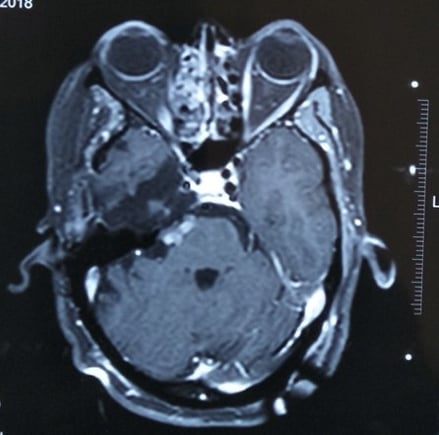
Trigeminal Schwannoma is a benign tumour of the nerve sheath arising from the perineural Schwann cells of the trigeminal nerve (5th cranial nerve). Trigeminal schwannomas are rare accounting for 0.07–0.3% of all intracranial tumours and 0.8%–5% of intracranial schwannomas. they usually present with facial pain, numbness and paraesthesia in the distribution of one or all the divisions of the trigeminal nerve depending on the location of the tumour. MRI is the gold standard for evaluation of trigeminal schwannomas because of its multiplanar capabilities and better soft tissue contrast. It helps in diagnosis and in planning the surgical approach. Surgery is usually performed to remove schwannomas, although radiosurgery is commonly used for schwannomas, particularly if they are small or have recurred after surgery

Residual tumour, which was then treated with Radiosurgery

Pre-operative MRI
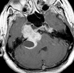
Vestibular schwannomas or acoustic neuromas are the most common and typically cause hearing loss and ringing in the ears (tinnitus); as they enlarge they may also cause imbalance and incoordination as well as facial weakness. They arise from the vestibular (8th cranial nerve). Large tumours need surgery to release the pressure on the brainstem, whilst smaller tumours could be treated with radiosurgery. These tumors are removed through a small skull opening behind the ear. Facial nerve, which is just adjacent to this tumor, is monitored during surgery.
2.VESTIBULAR NERVE SCHWANOMA (acoustic neuromas)
MRI
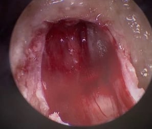
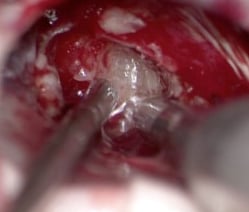
Tumour removal
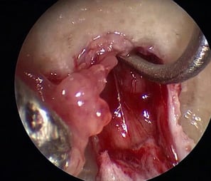
Intraoperative facial nerve monitoring
Use of endoscopy during surgery
Complete tumour removal with intact facial nerve


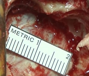

Mini Craniotomy

3.GLOSSOPHARYNGEAL NERVE (9th nerve)
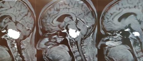
This behind tumor originates from the 9th(glossopharyngeal) nerve and usually compress adjacent nerve(10th&11th) and brain stem which monitor vital function like blood pressure & respiration. This 50-year-old lady had severe headaches, imbalance while walking, and difficulty in swallowing and speaking.
The MRI brain showed a large tumour in front of the brainstem causing severe compression and dysfunction of the nerves involved in swallowing. The patient did very well after surgery.
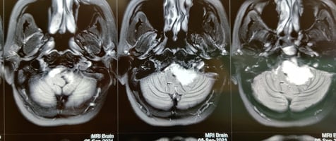

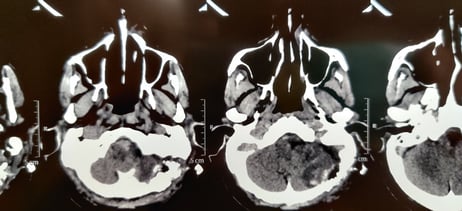

MRI-Before surgery
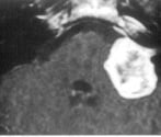
CT Scan-After surgery
MRI-Before surgery

© Copyright2024 - Dr Ashok Hande - All rights reserved
Clinic Details
Timings: 11 AM - 8 PM
Fortis Hospital
Contact Details for Emergency(24 Hrs)